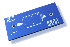
Sensor Chips and Disposable Cartridges
Low-cost and disposable sensor chips and microfluidic cartridges (patent pending) compatible with IRIS platform. Manufacturing using standard Si processing techniques provides superb quality control ensuring repeatability and scalability.
How it works?
Interferometric Reflectance Imaging Sensor, IRIS – builds on strong lineage of basic and applied research at Boston University and offers high-sensitivity and quantitative detection through an approach defying the conventional wisdom. Instead of enhancing the signal through optical resonances, we exploit the power of signal averaging in shot noise limited operation to achieve virtually unlimited sensitivity in a simple interferometric platform.
IRIS technique also overcomes the Achille’s Heel of label free detection: the bulk effect – the background signal due to concentration and temperature variations in the analyte solution.
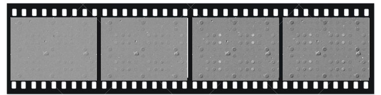
Real-time visualization of the microarray on the sensor surface provide intuitive monitoring of binding experiments.
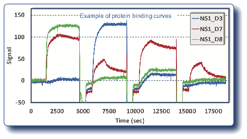
Multiplexed binding curves are extracted from the images acquired in real-time. Antibodies with unique binding characteristics for dengue NS1 are shown here (*). Rapid determination of the serotype specificity for each antibody will aid in the creation of serotype specific immunoassays.
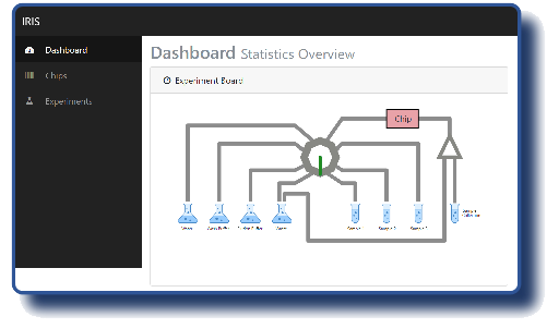
What does IRIS platform offers ?
A compact instrument with 12” X 18” footprint including all fluidics.
- Electronic controller integrated with on board computer.
- Platform independent user interface, run experiments on your iPad
- Fully automated experiments.
- 8-channel fluidics.
- Small dead volume.
- Removable fluidic tray.
- Fluidics with flexible configuration.
- Future expansion possibility for high throughput.
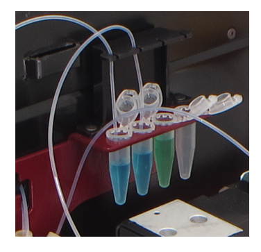
IRIS is configured to have four Eppendorf tubes for samples and four 250ml bottles for wash, buffer and waste. The minimum sample volume is 100 µl and can be further reduced. Automatic fluid handling allows for collecting and saving a sample. In addition, Flowrate can be adjusted across a large range using a syringe pump.
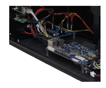
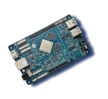
Custom electronic controller is integrated with an on-board computer for image acquisition and processing. Its User interface is platform independent and can be accessed via a web browser on local WiFi network. The device is also compatible with handheld devices, so it is convenient to run experiments using your iPad and check the status without leaving your desk.
- Intuitive graphic user interface to plan experiments.
- User configurable binding experiments.
- Automatic spot finding compatible with .gal files.
- Real-time image analysis and curve generation.
Quantified Measurements:
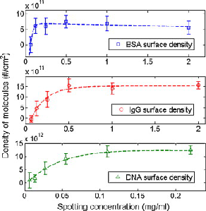
We have quantitatively calibrated the interferometric sensing to the surface-bound concentrations and masses of adsorbed layers of nucleic acids and proteins allowing us to calibrate the measured height to the absolute density of surface-bound molecules. IRIS has a linear dynamic range of nearly 4 orders of magnitude, which is comparable to standard SPR and considerably better than multiplexed alternative (SPRi) (*).
Applications
We cover the entire range of biological analytes including proteins, nucleic acids, peptides, and small molecules
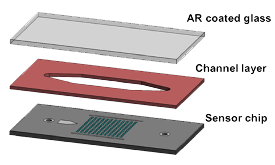
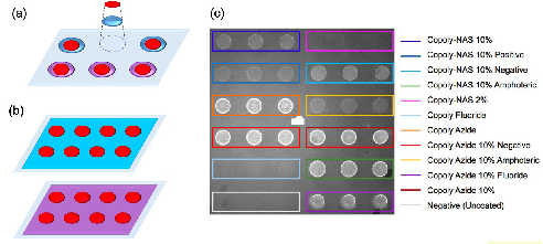
Si chips provide a large sensor area (5x7mm) and guide markings for robotic spotters for printing a microarray of ligands. Reference regions and rulers enable automated image analysis and quantification. Easy and intuitive assembly of disposable cartridges at room temperature protects the biological activity of immobilized ligands.
Oxide surface provides compatibility with glass surface chemistries. Multiple surface chemistry coatings can be used on the same IRIS chip simultaneously. IRIS allows the user to quickly understand which surface chemistry would better suit any specific ligand/analyte combination. Furthermore, IRIS eliminates the limitation regarding the number of molecules with different chemistries that can be immobilized on the same support.
Examples with small MW analytes:
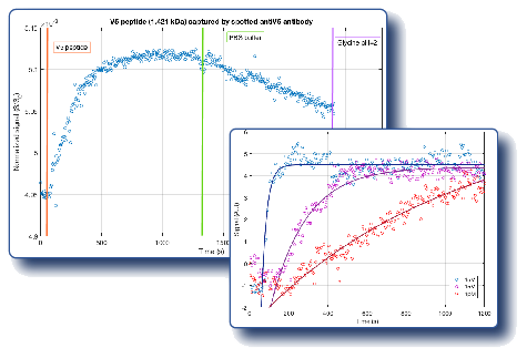
We have demonstrated high signal-to-noise ratio measurements of 1.4 kDa V5 peptide binding to immobilized antibodies. These results display average signal from 60 spots indicating >10-plex capability for peptide and small molecule analytes. Biotin (244 Da) at analyte concentrations of 1pM, 1nM and 1µM binding to immobilized avidin validates the high sensitivity.
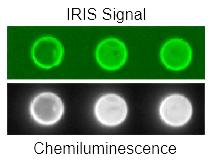
Compatible with Fluorescence and Chemiluminescence:
A dual detection system for protein arrays that combines IRIS with chemiluminescence for hepatitis B surface antigen has been developed. The dual detection system allows to combine the analytical capability of optical interference detection with the established clinical utility of chemiluminescence detection (*). IRIS chips enhance fluorescence intensity by optical interference (†).
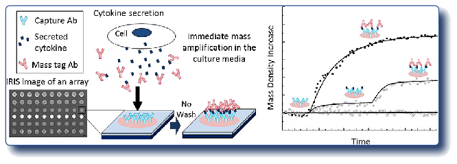
Dynamic Detection of Cytokine Secretion:
Monitoring cytokine release by cells allows the investigation of cellular response to specific external stimuli, such as pathogens or candidate drugs. We demonstrated a quantitative dynamic detection of interleukine-6 (IL-6), a pro-inflammatory cytokine and “mass tags” can be used concurrently with the target analyte to eliminate an additional wash and binding step. Successful label-free detection of IL-6 in cell culture medium (with 10% serum) has been reported (‡).